
- Heart Failure
- What is heart failure?
- What causes it?
- How is it diagnosed?
- Heart failure with preserved ejection fraction (HFpEF)
- How do we treat it?
- Prevention
- Helpful Links
Heart Failure
The heart pumps blood into the lungs (pulmonary circulation) and the rest of the body (systemic circulation) simultaneously. When healthy, the heart has the capacity to not only meet the blood flow needs of critical organs like the brain, kidneys and liver but also to increase its output to meet the changing needs of the body such as during exercise, mental stress or illness.
What is heart failure?
Heart failure occurs when the heart is unable to meet the blood flow requirements of the tissues. The heart may fail to contract with normal power (measured as the left ventricular ejection fraction), initially under stress and later at rest. This is called systolic heart failure, or heart failure with reduced ejection fraction (HFrEF). The other type – heart failure with preserved ejection fraction (HFpEF), previously called diastolic heart failure – is due to stiffness of the heart muscle, which prevents the heart from filling normally. It is now the most common form of heart failure, is closely linked to hypertension, obesity, diabetes, atrial fibrillation and increasing age, and is frequently missed because the resting echocardiogram often looks normal. Our HFpEF assessment program is described in the HFpEF tab.
The body adjusts to heart failure in a number of ways which partly compensate, but as the disease progresses exercise capacity falls, and salt and water accumulate in the lungs (pulmonary oedema) and the rest of the body, causing worsening breathlessness, liver congestion, swelling of the abdomen and legs, and sleep disruption (central sleep apnoea). Initially, people only notice these changes when exerting themselves and often think it is part of “ageing” or being unfit, only seeking help when their shortness of breath becomes severe.
Some patients have a combination of both systolic and diastolic heart failure. Heart failure of either type is a serious condition which impacts on length and quality of life, but fortunately there are many treatments and lifestyle changes that can improve both survival and quality of life.
What causes it?
Heart muscle damage can be caused by various factors. The commonest causes are coronary artery disease (‘heart attack’) and cardiomyopathy, or heart muscle disease. Genes play an important role in cardiomyopathy and in coronary disease, diabetes and hypertension.
Hypertension leads to heart muscle thickening (hypertrophy) and therefore stiffness. It is a common cause of heart failure with preserved ejection fraction (HFpEF), as are obesity, diabetes and atrial fibrillation. Reduced contraction occurs late in the disease.
Heart valve disease leads to narrowed (stenosis) or leaking (regurgitation) valves – most commonly the aortic or mitral valves are involved. This typically occurs gradually but can change suddenly, such as when a valve is infected (endocarditis). Viral infections can affect the heart muscle (myocarditis). Rheumatic fever is now uncommon in Australia outside Aboriginal and Torres Strait Islander communities.
Heart muscle can also be damaged by drugs and toxins, in particular alcohol, but also cancer drugs (anthracyclines or trastuzumab, also known as Herceptin).
Obstructive sleep apnoea is increasingly recognised as a cause of heart failure.
Heart rhythm disorders can cause or worsen heart failure – atrial fibrillation, atrial flutter and, less commonly, supraventricular tachycardia (see Heart Rhythm Disorders).
How is it diagnosed?
Heart failure is diagnosed by a combination of symptoms (breathlessness, abdominal or ankle swelling), signs of fluid overload in the chest, abdomen and legs when examined, and confirmed by evidence of abnormal heart function (either reduced contraction or preserved contraction with markedly increased stiffness). This evidence is usually obtained using cardiac ultrasound (echocardiogram). When the ejection fraction is preserved and the resting scan looks normal, the diagnosis often depends on testing the heart during exercise – see the HFpEF tab.
Dilated cardiomyopathy: the heart is enlarged and its function reduced.
Hypertrophic cardiomyopathy: the heart is not enlarged but is stiff – the muscle is markedly thickened and the cavity smaller.
A blood test for natriuretic peptides (BNP or NT-proBNP) is now a standard part of diagnosis. A normal level makes heart failure unlikely; a raised level supports the diagnosis and helps guide treatment.
Other tests used to determine whether heart failure is present, and its cause, include blood tests, chest x-ray, coronary angiography, CT scans and nuclear scans. Cardiac MRI is used when the cause is unclear – to look for scar, inflammation (myocarditis) or infiltrative disease.
Related: Invasive Cardiac Diagnostics · Invasive haemodynamic assessment
Heart failure with preserved ejection fraction (HFpEF)
HFpEF is now the most common form of heart failure, yet it is frequently missed. In many patients the resting echocardiogram looks normal, and the diagnosis is only revealed when the heart is assessed under load.
Central Sydney Cardiology, working with the University of Sydney, offers one of the most comprehensive HFpEF assessment programs in the country. Our approach moves beyond a single resting scan: where appropriate, we assess the heart across rest and exercise, and at the level of its underlying biology.
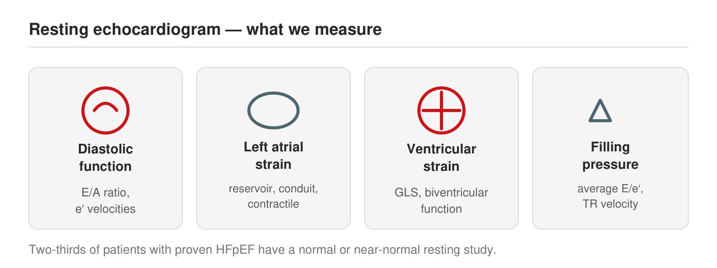
At rest we assess diastolic function, left atrial size and strain, biventricular strain, and estimated filling pressures. In many patients these are normal at rest, which is why exercise assessment is central to the diagnosis.
The resting study
Comprehensive baseline phenotyping
Every assessment begins with a detailed resting echocardiogram that goes well beyond ejection fraction: diastolic function, left atrial volume and reservoir strain, biventricular global longitudinal strain, and estimated filling pressures. This establishes the baseline and identifies the roughly one-third of patients whose HFpEF is already evident at rest. For the majority, whose resting study looks normal or borderline, the next step is assessment under load (O’Sullivan, Wilcox et al, Eur J Heart Fail 2026).
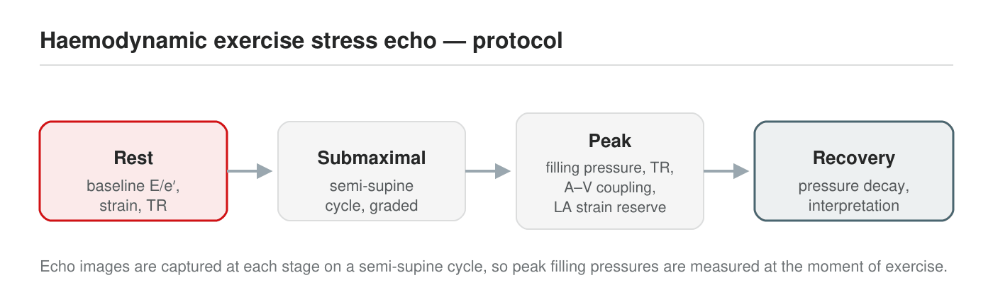
Our haemodynamic exercise protocol uses semi-supine cycle ergometry, allowing echocardiographic measurements to be captured at true peak exercise, when abnormal filling pressures and impaired cardiac reserve are unmasked.
Exercise and advanced assessment
Testing the heart under load
- Haemodynamic exercise echocardiography to unmask abnormal filling pressures and impaired cardiac reserve that are invisible at rest.
- Cardiopulmonary exercise testing (CPET) to objectively quantify exercise capacity and identify the cardiac and non-cardiac contributors to breathlessness.
- Invasive exercise right heart catheterisation, the diagnostic gold standard, for patients in whom the diagnosis remains uncertain.
- Molecular and metabolic profiling, linking the clinical picture to the underlying cardiometabolic drivers of disease.
This depth of phenotyping allows a precise diagnosis and a treatment plan tailored to each patient’s specific disease pathway, rather than a one-size-fits-all approach.
Research and clinical trials
Patients assessed through the program may be eligible for clinical trials led by Professor John O’Sullivan, including the investigator-initiated pEF-NAD, CardioNAD and pEF-EX studies. The program is also establishing access to novel industry-sponsored HFpEF trials, giving patients an early route to emerging therapies alongside expert clinical care.
How do we treat it?
Modern heart failure management is a team-based approach which includes the patient’s family, family doctor, specialist heart failure nurses and physiotherapists (heart failure exercise programs), and is community, rather than hospital, based. A key strategy is to detect early deterioration of heart failure (decompensation) so that treatment can be adjusted and hospitalisation avoided.
The key elements are lifestyle changes, drugs, treatment of sleep apnoea and devices.
Lifestyle changes include smoking cessation, dietary changes (a low-salt diet, alcohol restriction and a limit on fluid intake when advised), weight reduction and tailored exercise programs.
Drug treatment improves quality and length of life, proven in many clinical trials. For heart failure with reduced ejection fraction, four classes of medication are started together or in quick succession and adjusted to the highest tolerated doses:
- A renin–angiotensin system inhibitor – sacubitril/valsartan (an ARNI) is preferred; otherwise an ACE inhibitor (perindopril, ramipril and others) or an ARB (irbesartan, candesartan and others).
- A beta-blocker (bisoprolol, carvedilol, metoprolol succinate, nebivolol).
- A mineralocorticoid receptor antagonist (spironolactone, eplerenone).
- An SGLT2 inhibitor (dapagliflozin, empagliflozin).
Diuretics or ‘water tablets’ (frusemide and others) are used when fluid overload (‘congestion’) occurs or is likely to. Ivabradine, which slows the heart rate, is added for some patients in normal rhythm whose heart rate stays high on a beta-blocker. Iron deficiency is common in heart failure and is treated with an iron infusion, which improves symptoms. Anticoagulants (‘blood thinners’ – apixaban, rivaroxaban or dabigatran, or warfarin) are needed when the risk of stroke is high, such as in atrial fibrillation.
For heart failure with preserved ejection fraction (HFpEF), an SGLT2 inhibitor reduces hospital admissions and cardiovascular death and is now PBS-listed for heart failure regardless of ejection fraction. Diuretics control congestion, and treating the conditions that drive HFpEF – high blood pressure, atrial fibrillation, diabetes, obesity and sleep apnoea – is central. Newer options, including weight-loss medicines for people with obesity, are improving symptoms in trials, and our HFpEF program can offer eligible patients access to clinical trials of emerging therapies (see the HFpEF tab).
Diagnosis and treatment of sleep apnoea matters. Obstructive sleep apnoea is treated in the usual way, with CPAP and weight loss. Central sleep apnoea in heart failure is different: mask ventilation with adaptive servo-ventilation increased mortality in a large trial of patients with reduced ejection fraction and is not used in that group; optimising the heart failure treatment itself is the first step.
Implanted devices have two basic functions: 1) to treat arrhythmias and 2) to treat heart failure.
Defibrillators (ICDs) reduce the risk of sudden death due to serious heart arrhythmias. They are used for primary prevention in those with systolic heart failure (reduced ejection fraction) and secondary prevention when a significant asymptomatic ventricular arrhythmia or cardiac arrest has occurred.
Biventricular pacing (cardiac resynchronisation) is a heart failure treatment which can improve the efficiency of the heart’s contraction by pacing both right and left ventricles simultaneously. These devices are usually defibrillators as well.
These devices can be used to detect changes in heart rhythm (AF) and lung water (thoracic impedance) which can predict impending heart failure decompensation.
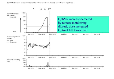
Country patient with ICD and worsening heart failure detected by remote monitoring
Prevention
Heart failure is a serious condition. Prevention and early detection are critical and have been the focus of major efforts to reduce the health burden of chronic heart failure by encouraging a healthier lifestyle and aggressively managing risk factors for heart failure (coronary artery disease, hypertension, obesity, diabetes, kidney disease, obstructive sleep apnoea).
Genetic testing can identify family members at risk of future heart failure due to inherited diseases like cardiomyopathy. A natriuretic peptide blood test in people at risk can identify heart failure earlier, when treatment has the greatest benefit.
The Heart Foundation has information on how to keep your heart healthy, prevent heart failure and manage heart failure if it develops.