
Invasive haemodynamic assessment measures the pressures and blood flow inside the heart and lungs directly, with thin catheters passed through a vein or artery. It answers questions that echocardiography and CT cannot settle: whether breathlessness is caused by high pressure in the lungs or by a stiff heart, how severe a valve problem really is when tests disagree, and whether the lining of the heart is constricting it. The study takes 30–60 minutes under local anaesthetic with light sedation. Sometimes it includes measurements before and after a fluid bolus, during exercise or after a test medication. Patients go home the same day. A/Prof Jordan Fulcher performs complex left and right heart studies at Royal Prince Alfred Hospital and Strathfield Private Hospital.
What the study measures
A right heart catheter records pressures in the right atrium, right ventricle and pulmonary artery, and the “wedge” pressure that reflects the left side of the heart. It measures how much blood the heart pumps each minute (cardiac output), can involve measurement of oxygen levels in each chamber to detect and quantify holes in the heart, and calculates the resistance of the lung circulation. A left heart catheter, passed through an artery at the wrist or groin, records pressures in the left ventricle and aorta and, when combined with echo, allows valve gradients and areas to be calculated. Measurements can be repeated during supine bicycle exercise, after a fluid load, or after a medication such as dobutamine or a pulmonary vasodilator, to unmask problems that are absent at rest.
When it is needed
- Pulmonary hypertension. Diagnosis requires a mean pulmonary artery pressure above 20 mmHg at catheterisation, and the study separates pressure caused by lung-vessel disease (pre-capillary) from pressure transmitted from a failing or stiff left heart (post-capillary) — a distinction that determines treatment. International guidelines require right heart catheterisation before pulmonary arterial hypertension is diagnosed or its treatment started (ESC/ERS 2022, Class I).
- Heart failure with preserved ejection fraction (HFpEF). Breathlessness with a normal pumping fraction is common and often unexplained by resting tests. A wedge pressure of 15 mmHg or more at rest, or 25 mmHg or more during exercise, confirms the diagnosis (ESC 2021).
- Valve disease when tests disagree. In aortic stenosis with a low gradient and reduced heart function, measurements during a dobutamine infusion distinguish true severe stenosis from a valve that only appears severe because flow is low. In mitral stenosis and regurgitation, exercise measurements clarify symptoms that do not match the resting echo. Right heart catheterisation is recommended before any tricuspid valve procedure, because echo underestimates lung pressures when the tricuspid valve leaks severely (ESC/EACTS 2025).
- Holes in the heart. Before an atrial septal defect is closed, the size of the shunt and the lung-vessel resistance are measured directly when the echo suggests raised pressures.
- Constriction versus restriction. A thickened pericardium squeezing the heart (constrictive pericarditis, which surgery can cure) and a stiff heart muscle (restrictive cardiomyopathy, which it cannot) look alike on imaging. Simultaneous pressure recordings from both ventricles during breathing separate them with high accuracy (Talreja et al, JACC 2008).
- Before structural procedures and transplant assessment. Haemodynamics guide suitability for TAVI, TEER and valvuloplasty, and are required in the work-up for advanced heart-failure therapies.
How the study is done
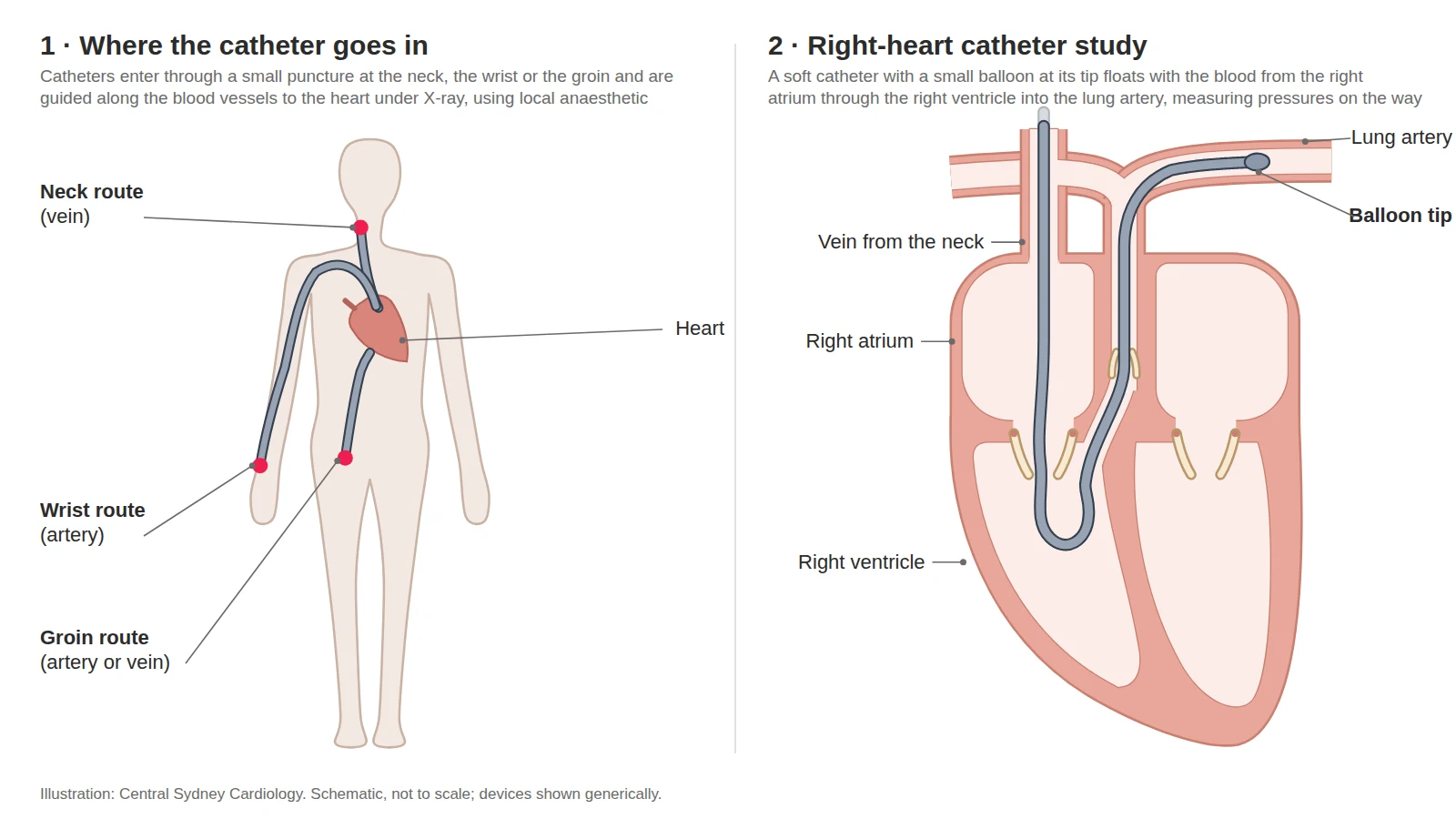
The study is performed in the cardiac catheterisation laboratory. After local anaesthetic, a small sheath is placed in a vein at the neck, arm or groin (for the right heart) and, when needed, in the artery at the wrist or groin (for the left heart). A balloon-tipped catheter is floated through the right heart into the pulmonary artery while pressures are recorded; you may feel brief palpitations as it passes through the ventricle. Blood samples may be taken for oxygen measurement. Cardiac output is measured by injecting cold saline (thermodilution) and by the Fick method from oxygen measurements — the two methods can disagree in a meaningful minority of patients, particularly with a leaking tricuspid valve or atrial fibrillation, which is why both are used when the classification matters (Hamilton, Fulcher et al, Pulm Circ 2025). If exercise measurements are needed you pedal a bicycle attached to the table while recordings continue. Coronary angiography is often combined with the study when valve intervention is being planned.
Risks
Right heart catheterisation carries a low complication rate. In a series of more than 7,000 procedures in patients with pulmonary hypertension, serious complications occurred in 1.1% — mostly access-site bleeding, temporary rhythm disturbance or a drop in blood pressure — and the procedure-related death rate was 0.055% (Hoeper et al, JACC 2006). Adding a left heart catheter and coronary angiography adds the small risks of arterial access, contrast reaction, kidney injury and, rarely, stroke or heart attack (under 1%). Injury to the pulmonary artery is very rare. Dobutamine can cause transient palpitations or chest tightness that settle when the infusion stops.
Recovery
Most studies are day procedures. After venous access you rest for one to two hours; after arterial access at the wrist a compression band is released over two hours, or after groin access you recover over four hours. You will be given the results at the follow-up consultation, together with the plan they support. Keep the puncture site dry for 48 hours; avoid heavy lifting for a few days; do not drive for 24 hours after sedation, or as advised.
For referring doctors
Who to refer. (1) Suspected pulmonary hypertension that needs confirming and classifying: a mean pulmonary artery pressure above 20 mmHg defines it; a wedge pressure of 15 mmHg or less with a pulmonary vascular resistance above 2 Wood units makes it pre-capillary; and a pressure–flow slope above 3 mmHg/L/min on exercise defines exercise pulmonary hypertension. Right heart catheterisation is Class I B before pulmonary arterial hypertension therapy is started (Humbert et al, Eur Heart J 2022). (2) Unexplained exertional breathlessness with a preserved ejection fraction and an intermediate probability of HFpEF on the HFA-PEFF or H₂FPEF score: an exercise right heart study showing a wedge pressure of 25 mmHg or more is the confirmatory test (McDonagh et al, Eur Heart J 2021). (3) Valve disease where the tests disagree: low-flow, low-gradient aortic stenosis (valve area 1.0 cm² or less with a mean gradient under 40 mmHg and a stroke volume index of 35 mL/m² or less) for dobutamine haemodynamics with assessment of flow reserve; mitral stenosis with symptoms out of proportion to the resting gradient (ACC/AHA 2020 Class 1 for exercise or invasive haemodynamics); severe tricuspid regurgitation before any intervention (ESC/EACTS 2025); and an ASD with suspected raised pulmonary vascular resistance before closure. (4) Suspected constrictive pericarditis versus restrictive cardiomyopathy: on simultaneous ventricular pressure recordings, a systolic area index above 1.1 identified surgically proven constriction with 97% sensitivity and 100% specificity (Talreja et al, JACC 2008). (5) Pre-transplant or LVAD haemodynamics at the request of the heart-failure team.
Work-up. Where available, please send the echocardiogram, ECG, spirometry or lung function, pathology and current medications. Studies are performed at RPA or Strathfield Private Hospital.
How to refer. HealthLink (EDI: centrasc), fax (02) 9336 2650, or email structuralheart@cardiology.sydney, marked to A/Prof Fulcher. Urgent: (02) 9336 2600, 08:00–18:00 Monday to Friday.
Haemodynamic assessment at Central Sydney Cardiology
A/Prof Jordan Fulcher performs right and left heart catheterisation at Royal Prince Alfred Hospital and Strathfield Private Hospital, and has published on the methods of cardiac output measurement and their effect on pulmonary hypertension classification (Pulm Circ 2025). Results are reviewed with the referring physician and, for valve and structural cases, at the RPA Heart Team meeting.
Frequently asked questions
Is a right heart catheter the same as an angiogram?
No. An angiogram images the coronary arteries with contrast dye through an artery. A right heart catheter passes through a vein to measure pressures and blood flow, usually without contrast. The two are often combined when both questions need answering.
Will I be awake?
Yes, with local anaesthetic and light sedation. Being awake matters for exercise studies, where you pedal a bicycle while measurements are taken.
How long will it take and can I go home the same day?
Thirty to sixty minutes, longer with exercise or drug testing. Almost all patients go home the same day after a few hours of observation.
Why do I need this if I have already had an echocardiogram?
Echocardiography estimates pressures indirectly and can be misleading — for example it underestimates lung pressures when the tricuspid valve leaks — and cannot measure pressures during exercise as reliably. Catheterisation gives direct measurements that decide treatment, such as whether pulmonary hypertension drugs are appropriate or whether a valve is severe enough to treat.
Is it covered by Medicare or my health fund?
Yes. Right and left heart catheterisation are Medicare-listed and covered by health funds.
Reviewed by A/Prof Jordan Fulcher, .