
For referring doctorsreferral criteria, work-up and how to refer
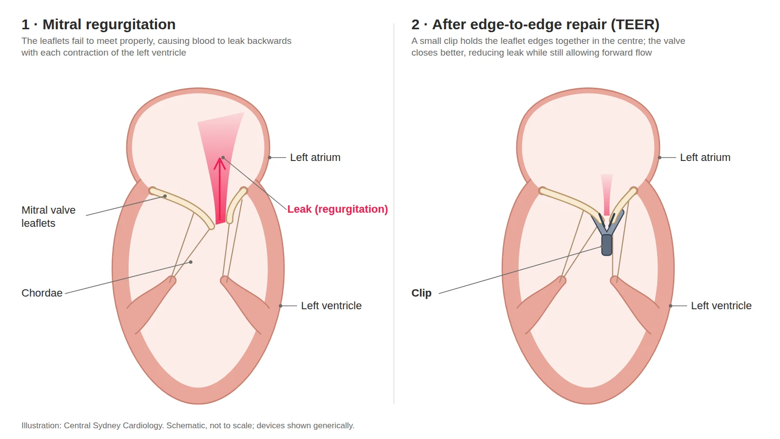
TEER (transcatheter edge-to-edge repair) treats a leaking mitral or tricuspid valve without open-heart surgery. A small clip — MitraClip, PASCAL or, for the tricuspid valve, TriClip — is delivered through a vein in the groin and fastened to the edges of the valve leaflets so that they close more completely. The procedure takes one to two hours under general anaesthetic and most patients go home after one or two nights. At Central Sydney Cardiology, A/Prof Jordan Fulcher assesses and works up all TEER candidates and performs TEER at Royal Prince Alfred Hospital.
What is mitral or tricuspid regurgitation?
The mitral valve sits between the left atrium and left ventricle; the tricuspid valve sits between the right atrium and right ventricle. Each should close tightly with every heartbeat. Regurgitation means the valve leaks, so blood flows backwards and the heart works harder. Mitral regurgitation is either primary (the valve itself is damaged — prolapse, a ruptured cord, degeneration) or secondary (the valve is structurally normal but is pulled apart by enlarged heart chambers, usually in heart failure). Tricuspid regurgitation is usually secondary, driven by atrial fibrillation, pulmonary hypertension or left-sided valve disease. Both cause breathlessness, fatigue and fluid retention, and both shorten life when severe. More on the conditions, diagnosis and treatment options is on our Mitral and Tricuspid Regurgitation page.
How TEER works
TEER is performed in the cardiac catheterisation laboratory under general anaesthetic, guided by transoesophageal echocardiography (an ultrasound probe in the oesophagus) and X-ray. A sheath is placed in the femoral vein at the groin under ultrasound guidance. For the mitral valve, a needle puncture is made through the thin wall between the two upper chambers to reach the left side of the heart; for the tricuspid valve no puncture is needed. The clip is positioned above the leak, opened, advanced across the valve and closed on the leaflet edges. Echocardiography then measures the remaining leak. If the position and result are satisfactory the clip is released. Sometimes a second or third clip may be added to reduce the leak further. Mitral TEER usually takes one to two hours; combined mitral and tricuspid procedures take longer. The technique adapts a long-established surgical stitch (the Alfieri repair); the first catheter procedure was performed in 2003 and more than 200,000 patients have been treated worldwide with MitraClip alone.
Who is TEER for?
TEER is considered in three situations:
- Severe primary mitral regurgitation when surgery is high risk. Surgical repair remains the first-choice treatment for most people with a damaged mitral valve because it removes the leak more completely. TEER is offered when the Heart Team judges surgical risk to be high — usually because of age, frailty, previous heart surgery or other illness — and the valve anatomy suits a clip (ESC/EACTS 2025 Class IIa; ACC/AHA 2020 Class 2a).
- Severe secondary mitral regurgitation in heart failure. For people whose leak is caused by a weakened, enlarged left ventricle, or enlarged left atrium, and whose symptoms persist despite optimised heart-failure medication (and a pacemaker-defibrillator where indicated), TEER added to medication reduces hospital admissions and improves survival and quality of life. The 2025 ESC/EACTS guideline gives this a Class I recommendation when specific criteria are met (ejection fraction 20–50%, left ventricular end-systolic diameter ≤70 mm, pulmonary pressure ≤70 mmHg, no severe right-heart failure).
- Severe tricuspid regurgitation. For people who remain symptomatic despite diuretics and are at high risk for surgery, tricuspid TEER improves symptoms and quality of life provided the right ventricle is not severely weakened and there is no fixed pulmonary hypertension (ESC/EACTS 2025 Class IIa).
Suitability is a Heart Team decision after transoesophageal echocardiography and right heart catheterisation. Valve anatomy matters: leaflet length, the size of the gap, calcification and the number of jets all influence whether a clip will hold and reduce the leak without narrowing the valve. Some patients are better served by surgery; some with advanced right-heart failure or fixed pulmonary hypertension will not benefit from any valve procedure.
Evidence
Secondary mitral regurgitation. In the COAPT trial (614 patients with heart failure and severe secondary MR), adding TEER to optimised medication reduced heart-failure hospitalisations from 67.9 to 35.8 per 100 patient-years (hazard ratio 0.53, 95% CI 0.40–0.70) and two-year mortality from 46.1% to 29.1% (HR 0.62, 95% CI 0.46–0.82) (Stone et al, NEJM 2018); the survival benefit persisted at five years (Stone et al, NEJM 2023). A second trial, MITRA-FR, enrolled patients with larger ventricles and less severe leaks and found no benefit (Obadia et al, NEJM 2018) — the reason careful patient selection is central to the guideline criteria above. RESHAPE-HF2 (2024) again showed fewer heart-failure hospitalisations or cardiovascular deaths (a combined outcome) with TEER (rate ratio 0.64, 95% CI 0.48–0.85) (Anker et al, NEJM 2024).
Primary mitral regurgitation. EVEREST II compared TEER with surgery in patients fit for either. Surgery removed the leak more completely — by five years 28% of TEER patients had needed surgery compared with 9% of surgical patients — while TEER had far fewer major complications in the first 30 days (15% vs 48%) (Feldman et al, NEJM 2011; JACC 2015).
Leak reduction with current devices. Across all patients treated, about 95% are left with a leak of moderate or less — over 90% in the US national registry (2014–19) and 97–98% in the latest-generation device study — and about 90% with mild or less (Mack et al, JACC 2021; EXPAND G4, JACC CI 2023; CLASP IID, JACC CI 2022).
Tricuspid regurgitation. TRILUMINATE (350 patients in the primary analysis, 572 at two years) showed that TEER reduced TR to moderate or less in 87% of patients versus 5% with medication alone, with a meaningful gain in quality of life; deaths were no different at one or two years, and heart-failure hospitalisations were lower by two years (Sorajja et al, NEJM 2023; Kar et al, Circulation 2025). The Tri.Fr trial (France and Belgium) found the same pattern: 74% of TEER patients improved on a combined clinical score at one year versus 41% on medication alone, with no significant difference in deaths or heart-failure admissions (Donal et al, JAMA 2025). The benefit of tricuspid TEER, on current evidence, is symptoms and quality of life rather than survival. Untreated significant TR is associated with roughly twice the mortality of no or mild TR, independent of pulmonary pressures (Wang, Fulcher et al, Eur Heart J 2019).
Risks
TEER avoids the chest incision, heart-lung machine and long recovery of open surgery, but it is a major cardiac procedure and no valve intervention is risk-free. Common and usually minor: bruising or bleeding at the groin, a sore throat from the echo probe, temporary rhythm disturbance, some residual leak, and (after mitral TEER) a small hole in the wall between the atria that usually closes by itself. Uncommon but serious: death within 30 days in 1–2% of patients in contemporary device studies and 3–4.5% in the US national registry, whose patients were older and more symptomatic; stroke in about 1%; the clip holding only one leaflet (about 1–2% for the mitral valve, higher — around 5–6% — for the tricuspid valve), which may need a further clip or surgery; bleeding around the heart needing drainage in about 0.5–1%; the clip coming loose in under 0.5%; emergency surgery in under 1%; and narrowing of the repaired valve (a mean gradient above 5 mmHg in about a quarter of registry patients). Risks also include kidney injury from contrast, infection, and injury to the leg vessels. Individual risk depends on anatomy, heart function and general health, and is discussed openly before any decision to proceed.
Recovery
You will lie flat for four to six hours while the groin puncture seals, then walk the same evening or the next morning. Most patients go home after one to two nights, often with an echocardiogram before discharge. You will take aspirin — sometimes with clopidogrel for the first weeks — while the heart’s own tissue grows over the clip; if you already take an anticoagulant for atrial fibrillation this usually continues instead. Do not stop these medications without speaking to your cardiologist. Keep the groin dry for 48 hours and avoid baths, swimming and heavy lifting for a week. Austroads’ Assessing Fitness to Drive advises not driving a private vehicle for four weeks after valve repair, including MitraClip and tricuspid clips; for commercial licences the non-driving period is three months. Your treating team will confirm your individual guidance. Most people notice easier breathing within days to weeks. Follow-up is with an echocardiogram at about one month, then at 6–12 months and annually. Because you have a valve implant, tell your dentist and other doctors, and keep up dental care to reduce the small risk of valve infection.
For referring doctors
Who to refer
- Primary (degenerative) mitral regurgitation. Severe with symptoms, in a patient who is a poor candidate for open surgery — age, frailty, previous heart surgery or other significant illness — or who declines surgery. Anatomical suitability is confirmed at Heart Team review.IIa B ESC/EACTS 2025 · 2a ACC/AHA 2020
- Secondary mitral regurgitation in heart failure. Ejection fraction 20–50% with moderate-severe or severe regurgitation still present, and symptoms, after at least three months of optimised heart-failure medication and CRT where indicated. COAPT-type criteria: left ventricular end-systolic diameter ≤70 mm, pulmonary artery systolic pressure ≤70 mmHg, no severe right ventricular dysfunction, and no coronary disease needing revascularisation.I A ESC/EACTS 2025
- Tricuspid regurgitation. Severe with symptoms despite diuretics, at high surgical risk, provided the right ventricle is not severely weakened and there is no fixed pulmonary hypertension.IIa A ESC/EACTS 2025
Refer early. Results are worse once right-heart failure, kidney impairment or liver congestion are established. When in doubt, refer for an opinion — suitability is a Heart Team decision after transoesophageal echocardiography and right heart catheterisation.
Work-up
Send with the referral, where available:
- Most recent transthoracic echocardiogram
- ECG
- Current medications with doses
- Pathology
We arrange transoesophageal echocardiography, left and right heart catheterisation, and Heart Team review.
Where and funding
- Consultations
- Central Sydney Cardiology, Newtown
- Work-up
- RPA or Strathfield Private Hospital
- Procedure
- RPA
- Funding
- Mitral and tricuspid TEER are performed at RPA for patients approved through a multidisciplinary case conference.
How to refer
- HealthLink
- centrasc (EDI)
- structuralheart@cardiology.sydney
- Fax
- (02) 9336 2650
- Phone
- (02) 9336 2600 Mon–Fri 08:00–18:00
- After hours
- RPA Hospital (02) 9515 6111 — ask for the cardiologist on call
Mark referrals to A/Prof Jordan Fulcher.
TEER at Central Sydney Cardiology
A/Prof Jordan Fulcher is a structural heart interventional cardiologist who trained in transcatheter valve therapy at the Austin Hospital, Melbourne, and Leeds General Infirmary, UK. He assesses and works up all TEER candidates at Central Sydney Cardiology in Newtown and performs mitral and tricuspid TEER at Royal Prince Alfred Hospital, where cases are reviewed by the RPA structural Heart Team of interventional cardiologists, cardiac surgeons, imaging cardiologists and anaesthetists. He is a co-author of a 70-study meta-analysis of tricuspid regurgitation and survival (Eur Heart J 2019).
Frequently asked questions
Is TEER the same as MitraClip?
MitraClip is one brand of TEER device; PASCAL is another, and TriClip is the version designed for the tricuspid valve. All three are approved for use in Australia. The device is chosen to suit your valve anatomy.
Is TEER as good as surgery?
It depends on the problem. For a damaged (primary) mitral valve in someone fit for surgery, surgical repair removes the leak more completely and lasts longer, so surgery is usually recommended. TEER had far fewer major complications in the first 30 days (15% versus 48% in EVEREST II) and is preferred when surgical risk is high. For secondary mitral regurgitation in heart failure, TEER plus medication has the strongest trial evidence, and a 2024 trial found it non-inferior to surgery at one year with fewer complications (MATTERHORN, NEJM 2024).
How long does the clip last?
The clip is a permanent implant; heart tissue grows over it within months. Most repairs are durable, but a leak can recur, and a further clip or surgery is sometimes needed — in EVEREST II about one in four TEER patients needed surgery within five years, most in the first six months.
Will I have a general anaesthetic?
Yes. TEER is performed under general anaesthesia because the procedure is guided by a transoesophageal echocardiogram probe.
Is TEER covered by Medicare or my health fund?
Mitral and tricuspid TEER are available for Medicare-eligible patients after a formal case conference at RPA.
When can I drive again?
Austroads advises not driving a private vehicle for four weeks after valve repair including MitraClip or tricuspid clips; for commercial licences the period is three months. Your team will confirm before discharge.
Reviewed by A/Prof Jordan Fulcher, .