
For referring doctorsreferral criteria, work-up and how to refer
- Overview
- Mitral regurgitation
- Tricuspid regurgitation
- Symptoms
- How valve leaks are diagnosed and graded
- Treatment
- For referring doctors
- Frequently asked questions
Overview
Regurgitation means a heart valve leaks. The mitral valve (left side) and tricuspid valve (right side) are the valves that most often leak, and significant leaks are common: moderate or severe mitral regurgitation affects about 9% of people aged 75 and over (Nkomo et al, Lancet 2006). Mild leaks are very common, usually cause no symptoms and need only periodic review. Severe leaks cause breathlessness, fatigue, swelling and atrial fibrillation, lead to heart failure, and shorten life — yet in a US community study only about one in seven people with moderate or severe isolated mitral regurgitation underwent mitral surgery (Dziadzko et al, Lancet 2018). Treatment options now range from surgical repair to keyhole clip repair (TEER). A/Prof Jordan Fulcher assesses valve function at Central Sydney Cardiology and performs TEER at Royal Prince Alfred Hospital.
Mitral regurgitation
Primary (degenerative) mitral regurgitation is a problem with the valve itself: a floppy or prolapsing leaflet, a ruptured supporting cord, rheumatic scarring, endocarditis or age-related degeneration. Secondary (functional) mitral regurgitation is a problem with the ventricle or atrium around a structurally normal valve — after a heart attack or in cardiomyopathy the ventricle enlarges and pulls the leaflets apart (“ventricular” secondary MR); in long-standing atrial fibrillation the atrium and valve ring stretch (“atrial” secondary MR). The distinction matters because the treatments differ. Untreated severe primary MR carries a five-year survival of about 58% against 78% expected, and timely surgery is associated with a large reduction in mortality (Enriquez-Sarano et al, NEJM 2005); severe secondary MR in heart failure is independently associated with about a 1.5- to 1.8-fold higher mortality, over and above the weak ventricle itself (Goliasch et al, Eur Heart J 2018).
Tricuspid regurgitation
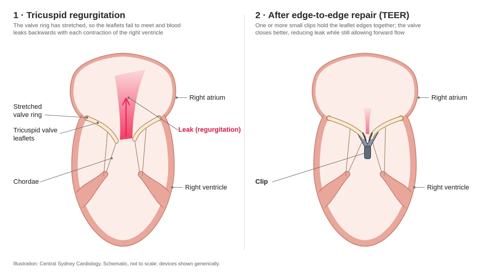
Tricuspid regurgitation is usually secondary — the right ventricle or the valve ring stretches because of left-sided valve disease, pulmonary hypertension, heart failure, atrial fibrillation or a pacemaker lead crossing the valve. Primary causes (endocarditis, rheumatic disease, injury, congenital abnormality) are less common. It was long dismissed as unimportant. It is not: across 70 studies and 32,000 patients, moderate or severe TR was associated with roughly double the risk of death, independent of pulmonary pressures and right ventricular function (Wang, Fulcher et al, Eur Heart J 2019), and in the Australian National Echo Database severe TR was found in 1.8% of all adults scanned and carried a 2.7-fold adjusted mortality risk (Offen et al, J Am Soc Echocardiogr 2022). Symptoms are fatigue, breathlessness, swelling of the legs and abdomen, and loss of appetite from liver congestion.
Symptoms
A leaking valve can be silent for years. Symptoms develop as the heart enlarges: breathlessness on exertion or lying flat, tiredness, palpitations (often atrial fibrillation), swollen ankles or abdomen, and reduced exercise capacity. Many people put a slow decline down to age. A new murmur, atrial fibrillation or unexplained breathlessness should prompt an echocardiogram.
How valve leaks are diagnosed and graded
Transthoracic echocardiography detects the leak and grades it by the width of the jet at the valve (vena contracta), the size of the leaking orifice and the volume of blood going backwards each beat. Mitral regurgitation is severe when the effective orifice is 40 mm² or more or the regurgitant volume 60 mL or more, and lower thresholds carry prognostic weight in secondary MR; tricuspid regurgitation is severe at an orifice of 40 mm² or a jet width of 7 mm, with “massive” and “torrential” grades beyond that. Transoesophageal echocardiography (a probe passed into the oesophagus under sedation) defines the mechanism and whether the valve can be repaired; cardiac MRI quantifies the leak and ventricular size when echo is uncertain; and right heart catheterisation measures lung pressures directly, which echo underestimates when the tricuspid valve leaks. Grading is repeated at intervals because leaks progress.
Treatment
Primary mitral regurgitation. Surgical repair — keeping your own valve — is the treatment of choice and is recommended for symptomatic severe MR, and for asymptomatic severe MR once the ventricle starts to enlarge or weaken (ejection fraction 60% or less, end-systolic diameter 40 mm or more), or when atrial fibrillation or raised lung pressure develops (ESC/EACTS 2025 Class I and IIa). Early repair in experienced centres carries a low risk (about 1% mortality) and durable results. For people at high surgical risk with suitable anatomy, TEER is the alternative (Class IIa).
Secondary mitral regurgitation. The first treatment is the ventricle: optimised heart-failure medication, and cardiac resynchronisation pacing where indicated, reduce the leak in about 40% of patients. If severe MR persists with symptoms, TEER added to medication reduces hospital admissions and improves survival and quality of life in patients meeting specific criteria (Class I A), on the strength of the COAPT trial (Stone et al, NEJM 2018). Surgery is reserved for those having bypass surgery anyway or who are unsuitable for TEER.
Tricuspid regurgitation. Diuretics control fluid but do not treat the valve. Repair at the time of left-sided valve surgery is recommended for severe (and considered for moderate) TR. Isolated severe TR is treated surgically in suitable patients, and by transcatheter repair or replacement in patients at high surgical risk who remain symptomatic on medication, provided the right ventricle is not severely weakened and there is no fixed pulmonary hypertension (Class IIa A) (Praz et al, Eur Heart J 2025). Treating TR earlier, before right-heart failure and kidney or liver congestion are established, gives better results.
For referring doctors
When to refer
- Any moderate or severe mitral or tricuspid regurgitation, for baseline assessment and a surveillance plan.
- All severe primary mitral regurgitation, including patients without symptoms, so that repair can be timed. An ejection fraction of 60% or less, or a left ventricular end-systolic diameter of 40 mm or more, is a Class I trigger for surgery; new atrial fibrillation, a pulmonary artery systolic pressure above 50 mmHg, or a dilating left atrium are Class IIa triggers.I / IIa ESC/EACTS 2025
- Heart failure with an ejection fraction below 50% and moderate-severe or severe secondary mitral regurgitation persisting after three months of optimised heart-failure therapy, with CRT where indicated — for TEER assessment when the COAPT criteria are met.I A ESC/EACTS 2025
- Symptomatic severe tricuspid regurgitation on diuretics, before right ventricular dysfunction, kidney impairment or liver congestion develop.
- New atrial fibrillation with any significant regurgitation.
Work-up
Send with the referral, where available:
- Echocardiogram report
- ECG and any rhythm monitoring
- Medication list, with heart-failure therapy doses
- Pathology
Where possible, please quantify the leak — effective orifice area, regurgitant volume, vena contracta — and report left and right ventricular size and function, the estimated pulmonary pressure, and left atrial volume.
We arrange transoesophageal echo, left and right heart catheterisation, and Heart Team review through our rooms.
Where
- Consultations
- Central Sydney Cardiology, Newtown
- Work-up
- RPA or Strathfield Private Hospital
- Procedure
- TEER is performed at RPA.
How to refer
- HealthLink
- centrasc (EDI)
- structuralheart@cardiology.sydney
- Fax
- (02) 9336 2650
- Phone
- (02) 9336 2600 Mon–Fri 08:00–18:00
- After hours
- RPA Hospital (02) 9515 6111 — ask for the cardiologist on call
Mark referrals to A/Prof Jordan Fulcher.
Frequently asked questions
I have mild mitral regurgitation. Is that serious?
No. Mild leaks are common, cause no symptoms and rarely progress quickly. An echocardiogram every few years is usually all that is needed.
Should a leaking valve be fixed before symptoms start?
For severe primary mitral regurgitation, often yes: repair is recommended once the ventricle begins to enlarge or weaken, or atrial fibrillation or raised lung pressures appear, because waiting for symptoms worsens the results.
Can a leaking valve be treated without surgery?
Yes, for suitable patients. TEER places a clip on the leaflets through a vein in the groin and is used for people at high surgical risk and for secondary mitral regurgitation in heart failure. Tricuspid TEER is available for symptomatic severe TR at high surgical risk.
Does tricuspid regurgitation need treatment?
Severe tricuspid regurgitation shortens life and causes fatigue, swelling and liver congestion. It is treated when symptoms persist despite diuretics, ideally before the right ventricle weakens.
When can I drive?
For a private licence, driving is unrestricted while you have no symptoms; commercial licence holders with significant valve disease need specialist review. Austroads advises against driving with symptoms on moderate exertion, and a four-week (private) or three-month (commercial) non-driving period after valve repair or replacement, including MitraClip and tricuspid clips.
Reviewed by A/Prof Jordan Fulcher, .