
For referring doctorsreferral criteria, work-up and how to refer
Mitral valvuloplasty — also called percutaneous mitral balloon commissurotomy (PMBC) or percutaneous transvenous mitral valvuloplasty (PTMV) — opens a narrowed mitral valve by inflating a specially shaped balloon inside it, without open-heart surgery. It is the treatment of choice for symptomatic rheumatic mitral stenosis when the valve anatomy is suitable. The procedure takes one to two hours, most patients go home the next day, and A/Prof Jordan Fulcher performs it at Royal Prince Alfred Hospital and Strathfield Private Hospital.
What is mitral stenosis?
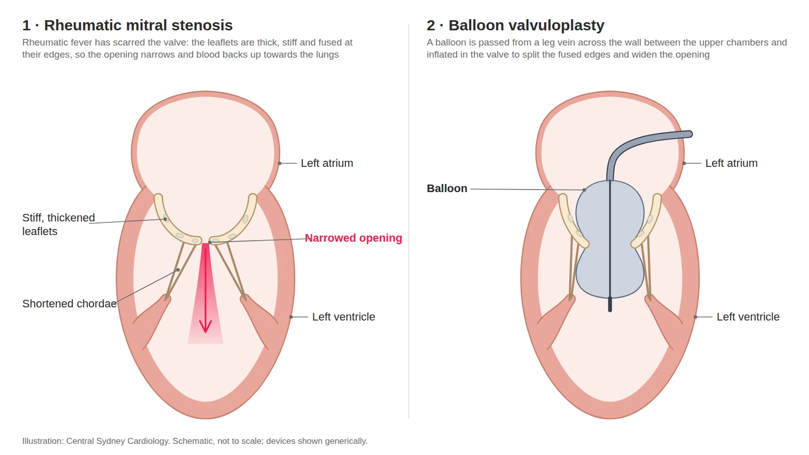
Mitral stenosis is narrowing of the valve between the left atrium and left ventricle. Worldwide its cause is almost always rheumatic heart disease — scarring that follows rheumatic fever, itself a reaction to untreated streptococcal throat or skin infection, usually in childhood. The valve leaflets thicken and fuse at their edges (the commissures), the opening shrinks and pressure builds up in the left atrium and lungs, causing breathlessness, fatigue, palpitations, atrial fibrillation and, if clots form, stroke. In Australia rheumatic heart disease is uncommon in the general population but remains a serious problem for Aboriginal and Torres Strait Islander peoples — more than 7,500 people were on rheumatic heart disease registers at the end of 2024, four in five of them First Nations people — and is also seen in people who grew up in the Pacific, Asia, Africa and the Middle East (AIHW 2025; Katzenellenbogen et al, J Am Heart Assoc 2020). Mitral stenosis is diagnosed and graded by echocardiography; a valve area of 1.5 cm² or less is clinically significant.
How mitral valvuloplasty works
The procedure is performed in the cardiac catheterisation laboratory under general anaesthetic with transoesophageal echocardiography. Sheaths are placed in the femoral vein and artery at the groin. A needle puncture through the wall between the atria gives access to the left atrium and the mitral valve. The Inoue balloon — an hourglass-shaped balloon used for this purpose for more than thirty years — is advanced across the valve and inflated in steps of increasing size; each inflation splits the fused commissures a little further. After each step the valve opening, the pressure gradient and any leak are measured by echo and pressure recordings, and the procedure stops when the opening is adequate without a significant new leak. The catheters are then removed and the punctures sealed.
Who is mitral valvuloplasty for?
Valvuloplasty is recommended for people with symptoms and clinically significant mitral stenosis (valve area ≤1.5 cm²) whose valve anatomy is favourable — pliable leaflets, fused commissures and little calcium, usually summarised by an echocardiographic (Wilkins) score of 8 or less (ESC/EACTS 2025 Class I; ACC/AHA 2020 Class 1). It is also recommended for symptomatic patients who are unsuitable or high risk for surgery even when the anatomy is less favourable, and it is considered in people without symptoms who are at high risk of clots or of sudden deterioration — for example a history of embolism, new atrial fibrillation, high lung-artery pressure, planned major surgery, or pregnancy or a wish to become pregnant. It can be performed during pregnancy, preferably after 20 weeks, when symptoms persist despite medication (ESC/EACTS 2025). Valvuloplasty is not suitable when there is clot in the left atrium, more than mild mitral leak, heavy calcification of both commissures, or other valve or coronary disease that itself needs surgery; those patients are referred for surgical valve replacement.
Evidence
Balloon commissurotomy has been the standard treatment for rheumatic mitral stenosis since the late 1980s and gives an adequate valve opening without significant leak in about 90% of procedures (Iung et al, Circulation 1999). Its effect is durable but not permanent: in a large French series, 56% of all patients were free of cardiovascular death, further intervention and limiting symptoms at ten years (Iung et al, Circulation 1999), and of those with a good initial result, 30% still had a good functional result at twenty years (Bouleti et al, Circulation 2012). In younger patients with pliable valves, results are better: 88% event-free at ten years in one series (Fawzy et al, Cardiology 2009). Older age, calcification, a higher echo score, atrial fibrillation and a smaller valve area after the procedure predict earlier re-narrowing. When re-stenosis occurs, a repeat valvuloplasty is often possible and gives a good immediate result in about 90% (Iung et al, Eur Heart J 2000); otherwise surgical valve replacement is the alternative.
Risks
Common and usually minor: groin bruising, a small increase in mitral leak, brief rhythm disturbance, and a small residual hole in the atrial septum that closes or causes no problem. Uncommon but serious, from large series of the Inoue technique: a severe new mitral leak in about 1–2% (which may need urgent valve surgery), bleeding around the heart needing drainage in about 0.2–0.8%, stroke or embolism in about 0.5%, and death in around 0.1–0.2% (Chen & Cheng, Am Heart J 1995; Arora et al, Catheter Cardiovasc Interv 2002). Contrast reactions, infection and leg-vessel injury are rare. Risk is lower with favourable anatomy and experienced operators, and is discussed before any decision to proceed.
Recovery
You will lie flat for four to six hours and usually stay one night with rhythm monitoring, then walk the next morning. Keep the groin dry for 48 hours; avoid baths, swimming and heavy lifting for a week. Mitral valvuloplasty is not listed separately in Austroads’ Assessing Fitness to Drive: do not drive for at least 48 hours after the procedure, or as advised by your team. Breathlessness usually improves within days. Medications are reviewed before discharge: anticoagulation continues if you have atrial fibrillation, and secondary prophylaxis against rheumatic fever (regular benzathine penicillin) continues where it has been prescribed. Follow-up is with an echocardiogram at 1 month, then annual review with echocardiography, because re-narrowing can develop over years. Keep up dental care and tell your dentist about your valve.
For referring doctors
Who to refer
- Rheumatic mitral stenosis with symptoms and a valve area of 1.5 cm² or less. Class I C where the patient is too high-risk for surgery even when the anatomy is less favourable.I B ESC/EACTS 2025 · I B ACC/AHA 2020
- No symptoms, but a high risk of clot or sudden deterioration. A previous embolism, dense spontaneous contrast in the left atrium, new or paroxysmal atrial fibrillation, a pulmonary artery pressure above 50 mmHg at rest, planned major non-cardiac surgery, or pregnancy or a wish to become pregnant.IIa C ESC/EACTS 2025
- Pregnancy — refer early. Heart failure develops in about a third of pregnancies when the valve area is under 1.5 cm². Valvuloplasty is recommended when symptoms or a pulmonary pressure above 50 mmHg persist despite beta-blockers and diuretics, preferably after 20 weeks.
Not suitable when
- There is clot in the left atrium
- There is more than mild mitral leak
- Calcification is heavy or bicommissural
- The commissures are not fused
- Other valve or coronary disease itself needs surgery
Work-up
Send with the referral, where available:
- Echocardiogram, with planimetered valve area and mean gradient
- ECG
- Medication list
- Renal function
- Rheumatic fever and prophylaxis history
We arrange transoesophageal echocardiography to exclude appendage thrombus and grade the commissures, coronary assessment where age or symptoms indicate, and right heart catheterisation when pulmonary pressures are raised.
Where and funding
- Consultations
- Central Sydney Cardiology, Newtown
- Work-up
- RPA or Strathfield Private Hospital
- Procedure
- RPA for public patients; Strathfield Private Hospital for private patients
- Funding
- Medicare and health funds cover the procedure.
How to refer
- HealthLink
- centrasc (EDI)
- structuralheart@cardiology.sydney
- Fax
- (02) 9336 2650
- Phone
- (02) 9336 2600 Mon–Fri 08:00–18:00
- After hours
- RPA Hospital (02) 9515 6111 — ask for the cardiologist on call
Mark referrals to A/Prof Jordan Fulcher.
Mitral valvuloplasty at Central Sydney Cardiology
A/Prof Jordan Fulcher performs percutaneous mitral balloon commissurotomy at Royal Prince Alfred Hospital and Strathfield Private Hospital, including for patients referred from New Caledonia and the Pacific, where rheumatic heart disease remains common. Assessment and follow-up are at Central Sydney Cardiology, Newtown.
Frequently asked questions
Is valvuloplasty a cure?
It relieves the narrowing, often for many years, but the rheumatic scarring process can continue and the valve may narrow again. Regular echocardiography picks this up early; a repeat valvuloplasty or valve surgery can then be considered.
Why not just replace the valve?
Valvuloplasty keeps your own valve, avoids open-heart surgery, and does not commit you to lifelong warfarin (needed for a mechanical valve) or to a tissue valve that wears out. Surgery is reserved for valves that are too calcified or leaky, or when other heart surgery is needed anyway.
Can it be done during pregnancy?
Yes. When symptoms persist despite medication, valvuloplasty is recommended in pregnancy, ideally after 20 weeks, with measures to minimise the radiation dose to the baby.
Is it covered by Medicare or my health fund?
Yes. Medicare and health funds cover it. Public patients are treated at RPA Hospital. Private patients can also be treated at Strathfield Private Hospital.
When can I drive again?
Valvuloplasty is not listed separately in Austroads’ driving standards. Do not drive for at least 48 hours after the procedure, or as advised; your team will confirm before discharge.
Reviewed by A/Prof Jordan Fulcher, .