
For referring doctorsreferral criteria, work-up and how to refer
A patent foramen ovale (PFO) is a flap-like opening between the two upper chambers of the heart that fails to seal after birth; an atrial septal defect (ASD) is a true hole in the same wall. Both can be closed without surgery using a device delivered through a vein in the groin. PFO closure is recommended for selected people under 60 who have had a stroke that is most likely explained by the PFO; ASD closure is recommended when the hole is enlarging the right side of the heart. The procedure takes about an hour under general anaesthetic, most patients go home the next day, and A/Prof Jordan Fulcher performs it at Royal Prince Alfred Hospital and Strathfield Private Hospital.
What are a PFO and an ASD?
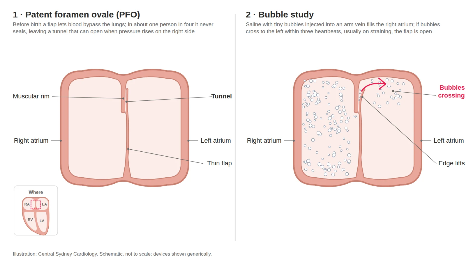
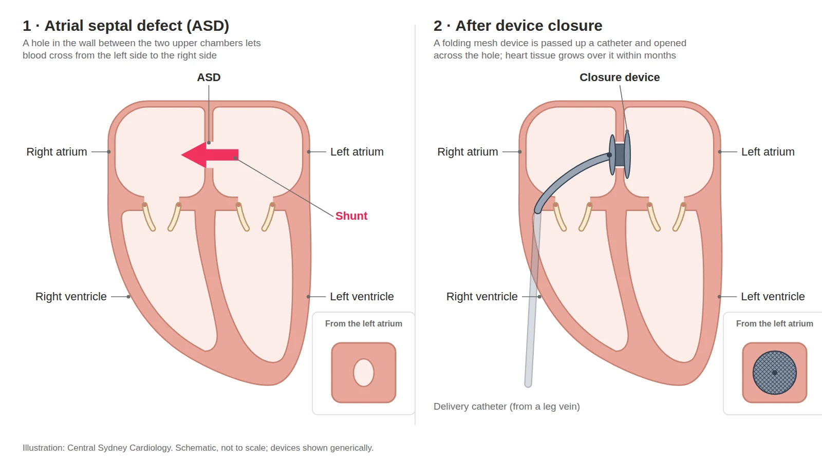
Before birth, blood bypasses the lungs through an opening between the right and left atria. In about one in four adults this opening never fully seals — a patent foramen ovale — and in most it causes no problem at all (Hagen et al, Mayo Clin Proc 1984). Occasionally a clot from a leg or pelvic vein can pass through a PFO to the arterial side and cause a stroke (“paradoxical embolism”). An ASD is different: a hole present from birth, usually in the middle of the wall (a secundum ASD, about 80% of ASDs). Blood shunts from left to right through it year after year. If the ASD is large enough, this flow can stretch the right atrium and ventricle and, in time, cause breathlessness, atrial arrhythmias, raised lung pressures and, rarely, paradoxical embolism. A PFO is usually diagnosed during an echocardiogram with a “bubble study” — saline microbubbles injected into a vein are seen crossing from the right atrium to the left through the wall between them. An ASD is usually seen on a transthoracic echocardiogram. Both PFOs and ASDs can be formally diagnosed with a transoesophageal echocardiogram.
How closure works
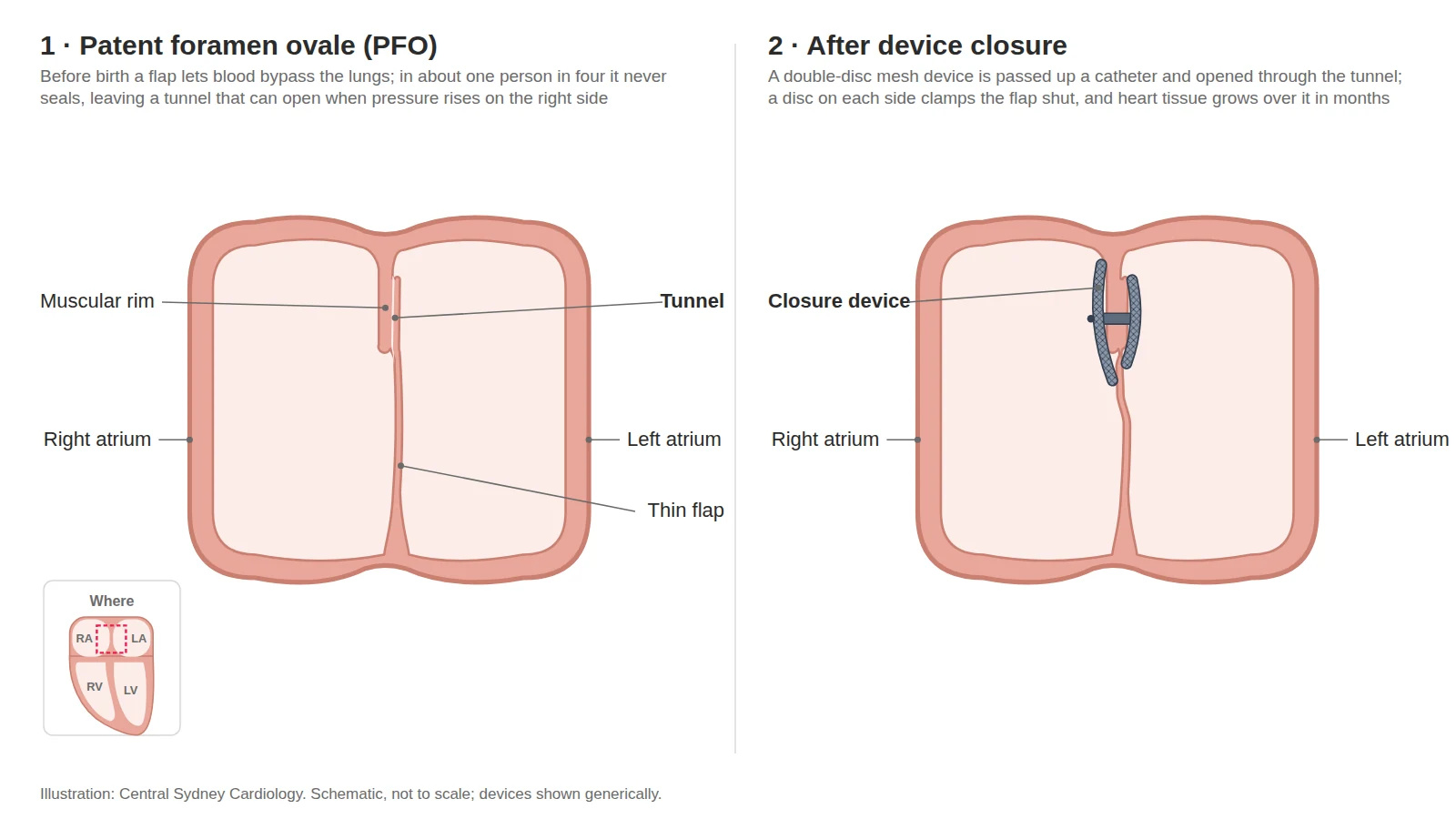
Closure is performed in the cardiac catheterisation laboratory, usually under general anaesthetic with transoesophageal echocardiography guiding device placement. A catheter is passed from the femoral vein into the right atrium and through the defect. The closure device — two fabric-covered discs joined by a short waist (Amplatzer Talisman PFO Occluder or Amplatzer Septal Occluder; other devices such as the Gore Cardioform suit particular anatomies) — is opened one disc on each side of the wall so that the hole is sandwiched shut. Its position and seal are checked by echo before release. The device stays permanently; the heart’s own tissue grows over it within months. The procedure takes about 45–90 minutes.
Who is closure for?
PFO closure is typically recommended for people aged under 60 who have had an ischaemic stroke for which no other cause can be found after thorough investigation and in whom the PFO is judged the likely cause — particularly when the PFO is large or accompanied by an atrial septal aneurysm. This is the position of the Australian and New Zealand Stroke Foundation guideline (strong recommendation), the Society for Cardiovascular Angiography and Interventions (2022) and the American Academy of Neurology (2020). Closure is not routinely recommended for migraine (three randomised trials did not meet their primary endpoints), for divers to prevent decompression illness, or for a PFO found incidentally without stroke. It may be considered for the rare platypnoea–orthodeoxia syndrome and in selected divers with prior decompression illness who wish to return to diving.
ASD closure is recommended, whether or not there are symptoms, when the shunt is large enough to enlarge the right heart and lung-artery pressures are not severely raised (ESC 2020 Class I). Device closure is the preferred method for secundum defects with adequate rims; other ASD types, very large defects and deficient rims need surgical closure. Closure after the age of 40 still relieves symptoms and protects the right heart, although atrial arrhythmias remain common in later life. Closure is not performed when lung-artery pressures are very high (Eisenmenger physiology).
Evidence
Pooling the six randomised trials of PFO closure after stroke (3,740 patients), closure reduced recurrent stroke from 1.09% to 0.47% per year (adjusted HR 0.41, 95% CI 0.28–0.60); the benefit was concentrated in people whose PFO was “probably” or “possibly” causal on the PASCAL classification, with no benefit when the PFO was an unlikely cause (Kent et al, JAMA 2021). In CLOSE, which enrolled only PFOs with an atrial septal aneurysm or a large shunt, there were no recurrent strokes after closure against 6% with antiplatelet tablets over five years (Mas et al, NEJM 2017); in REDUCE the hazard ratio was 0.23 (95% CI 0.09–0.62) at a median of 3.2 years and 0.31 (95% CI 0.13–0.76) at five years, when 25 patients needed to be treated to prevent one stroke (Søndergaard et al, NEJM 2017; Kasner et al, NEJM 2021). There is a small increased risk of new atrial fibrillation after closure — about four-fold, roughly 5% versus 1%, with about half of episodes transient in the first six weeks (Kent et al, JAMA 2021) — and a procedural complication rate of about 2% (Turc et al, JAHA 2018). For ASD, device closure has replaced surgery for most secundum defects, with major peri-procedural complications in about 1.5% in pooled case series (Abaci et al, Catheter Cardiovasc Interv 2013; Baumgartner et al, Eur Heart J 2021).
Risks
Common and minor: groin bruising, sore throat or headache after the echo probe, palpitations or brief rhythm disturbance. Uncommon but serious (about 1–2% in total): the device moving out of position (about 0.5%, usually retrieved by catheter, occasionally needing surgery), bleeding around the heart, stroke or air embolism, and new atrial fibrillation, which is usually temporary. A rare late complication of ASD devices is erosion through the heart wall (about 0.1–0.3%), which can occur months to years later and is why new chest pain or breathlessness should always be reported. Contrast reactions, infection and nickel sensitivity are rare. Individual risk is discussed before any decision to proceed.
Recovery
You will lie flat for four to six hours and usually stay one night, with an echocardiogram before discharge. Aspirin is taken for at least six months and clopidogrel may be prescribed for the first one to three months while tissue covers the device. Keep the groin dry for 48 hours; avoid baths, swimming and heavy lifting for a week. Austroads’ Assessing Fitness to Drive advises not driving a private vehicle for two weeks after a percutaneous procedure for a congenital heart lesion; for commercial licences the period is four weeks. A follow-up echocardiogram at four to six weeks and again at six to twelve months checks the device position and for any residual shunt. Tell your dentist and other doctors about the device; your cardiologist will advise whether antibiotic cover is needed for dental work in the first six months.
For referring doctors
Who to refer — PFO
- Aged 18–60 with a cryptogenic stroke. An ischaemic stroke, or an embolism to another artery, with no cause found and a PFO on bubble study — once the stroke work-up has excluded other causes: imaging of the brain and neck arteries, prolonged rhythm monitoring for atrial fibrillation, and clotting tests where indicated.2a AHA/ASA 2021 · Stroke Foundation living guideline
- Over 60 — case by case, not routine. AHA/ASA 2021 advises closure should rarely be performed in older patients, given limited trial data and more procedural complications.
- Known atrial fibrillation argues against closure. It is the likelier cause of the stroke.
The ANZ consensus 2026 advises a transcranial Doppler bubble study for every cryptogenic stroke at 60 or under, transoesophageal echo to confirm before closure, the PASCAL classification rather than the RoPE score alone to judge whether the PFO is the cause, and a joint neurology–cardiology decision.
Who to refer — ASD
- Any adult with an ASD and one of the following. An enlarged right atrium or ventricle on echo, a significant shunt (Qp:Qs of 1.5 or more), symptoms, an atrial arrhythmia, or a paradoxical embolism.
- Closure is recommended when the right heart is volume-loaded and pulmonary vascular resistance is below 3 Wood units, whether or not there are symptoms. Device closure is the method of choice for suitable secundum defects I C.I B ESC 2020
- Raised pulmonary pressures on echo need invasive haemodynamic assessment before any decision.
Work-up
Send with the referral, where available:
- Echocardiogram
- Stroke imaging and neurology correspondence
- ECG and rhythm monitoring
- Medication list
- Renal function
We arrange transoesophageal echo to define anatomy, rims and shunt; right heart catheterisation where pulmonary pressures are raised; and CT if needed.
Where and funding
- Consultations
- Central Sydney Cardiology, Newtown
- Work-up
- RPA or Strathfield Private Hospital
- Procedure
- RPA for public patients; Strathfield Private Hospital for private patients
- Funding
- Medicare and health funds cover transcatheter ASD or PFO closure “in a patient with documented evidence of right heart overload or paradoxical embolism”.
How to refer
- HealthLink
- centrasc (EDI)
- structuralheart@cardiology.sydney
- Fax
- (02) 9336 2650
- Phone
- (02) 9336 2600 Mon–Fri 08:00–18:00
- After hours
- RPA Hospital (02) 9515 6111 — ask for the cardiologist on call
Mark referrals to A/Prof Jordan Fulcher.
ASD and PFO closure at Central Sydney Cardiology
A/Prof Jordan Fulcher performs transcatheter ASD and PFO closure at Royal Prince Alfred Hospital and Strathfield Private Hospital. Assessment and follow-up are at Central Sydney Cardiology, Newtown.
Frequently asked questions
I have a PFO but have never had a stroke. Should it be closed?
No. A PFO is present in about a quarter of adults and, on its own, is not a reason for closure. Closure is recommended after a stroke that is most likely explained by the PFO, and in a few rare conditions.
Does PFO closure prevent migraine?
Three randomised trials did not show a convincing benefit, so closure is not recommended for migraine, even though some people with migraine with aura do have a PFO.
Will I still need blood thinners after closure?
Aspirin for at least six months while tissue grows over the device. Long-term anticoagulation is not needed for the device itself; if you have another reason for anticoagulation (such as atrial fibrillation) that continues.
Is the procedure covered by Medicare or my health fund?
Yes for eligible patients — Medicare and health funds cover transcatheter ASD or PFO closure “in a patient with documented evidence of right heart overload or paradoxical embolism”. Public patients are treated at RPA Hospital. Private patients can also be treated at Strathfield Private Hospital.
Can the device be felt, and will it set off airport security?
No. The device is small, inside the heart, and cannot be felt. Its nickel-titanium frame does not set off airport detectors, and current devices are MR-conditional, so an MRI can be performed under the conditions specified for the device. You will be given a device card to carry.
Reviewed by A/Prof Jordan Fulcher, .